Manufacturer: Revvity
Model Name: Opera Phenix Plus High-Content Screening System
Equipment Overview:
The Opera Phenix Plus is an automated High-Content Screening/Imaging system for quantitative fluorescence imaging of cells and complex 2D/3D biological models in multiwell plates. This system supports spinning-disk confocal imaging, simultaneous two-channel acquisition, high-sensitivity sCMOS detection, onboard liquid handling, environment control, and Harmony-based image acquisition and analysis with AI-powered analysis for high-throughput phenotypic screening workflows. - High-content imaging for quantitative analysis of 2D and 3D cell models - Multi-mode imaging: spinning-disk confocal, widefield, brightfield, and digital phase contrast, with simultaneous two-channel acquisition - Dual-camera parallel acquisition for efficient high-throughput imaging - Fully automated imaging workflow with Cytomat integration and onboard environmental control - Harmony software with machine learning and AI-based tools for segmentation and phenotypic analysis 👉 Value proposition: Fast, sensitive, and scalable imaging for complex biological models and large datasets.
Applications:
Designed for drug discovery, translational research, and advanced cell biology: - High-content and phenotypic screening Multiparametric analysis of cellular responses to compounds - 3D biology and complex models Imaging of Organoids, spheroids, and thick tissue imaging - Drug discovery and development Target validation, mechanism-of-action, toxicity assessment - Functional genomics CRISPR and RNAi-based screening assays - Live-cell and kinetic assays Calcium flux, cell dynamics, and time-lapse imaging - Cell painting and multiplex imaging High-dimensional morphological profiling
Instrument Overview:
The Opera Phenix Plus is an automated High-Content Screening/Imaging system for quantitative fluorescence imaging of cells and complex 2D/3D biological models in multiwell plates. This system supports spinning-disk confocal imaging, simultaneous two-channel acquisition, high-sensitivity sCMOS detection, onboard liquid handling, environment control, and Harmony-based image acquisition and analysis with AI-powered analysis for high-throughput phenotypic screening workflows. - High-content imaging for quantitative analysis of 2D and 3D cell models - Multi-mode imaging: spinning-disk confocal, widefield, brightfield, and digital phase contrast, with simultaneous two-channel acquisition - Dual-camera parallel acquisition for efficient high-throughput imaging - Fully automated imaging workflow with Cytomat integration and onboard environmental control - Harmony software with machine learning and AI-based tools for segmentation and phenotypic analysis 👉 Value proposition: Fast, sensitive, and scalable imaging for complex biological models and large datasets.
Technical features and specifications:
Imaging and Optics: - Spinning disk confocal imaging with high-sensitivity optics - Multi-mode imaging (confocal, widefield, brightfield, digital phase contrast) - High-resolution z-stack imaging for 3D samples Detection and Speed: - 16-bit sCMOS detection with parallel multi-camera acquisition - Simultaneous multi-channel fluorescence imaging - High-speed acquisition for high-throughput screening workflows (up to 105 fps with one camera at reduced frame size) Light Sources: - Laser lines: 405, 488, 561, 640 nm Objectives: - Water immersion objectives: 20x (NA 1.0, WD 1.7 mm), 40x (NA 1.1, WD 0.62 mm), and 63x (NA 1.15, WD 0.6 mm) Throughput and Automation: - Compatible with 96- and 384-well plates (and higher formats) - Automated imaging with motorized stage control and Cytomat integration - Designed for high-throughput screening workflows Environmental Control: - Temperature, CO₂, and humidity control for live-cell imaging Software and Analysis: - Integrated Harmony software for image acquisition and analysis - Machine learning and AI-assisted tools for image segmentation and phenotypic profiling - Data management, feature extraction, and phenotypic classification
Charges:
Please email us at earo@eddc.sg
Additional Info:
- (Autonomous use) Standalone for flexible, routine imaging - (Autonomous use) With robotic arm & Cytomat 5C +/- Opera Phenix for automated incubation & plate handling - (Staff Assisted) With robotic arm & Cytomat 5C and liquid handler integration +/- Opera Phenix for automated assay preparation
Equipment URL:
https://www.revvity.com/sg-en/product/opera-phenix-plus-system-hh14001000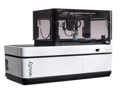
Other Equipment from EDDC
Loading...